
Chamber enlargement
Right atrial enlargement
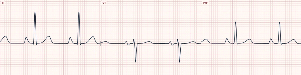
An enlarged right atrium produces a tall, peaked P wave in the inferior leads, while the P-wave width stays normal.
What the heart is doing
The right atrium depolarizes first, with forces pointing downward and forward. More right atrial muscle enlarges this early component, which stacks on top of the left atrial part.
Why it looks this way
This is right atrial enlargement because the P wave in lead II is peaked and taller than 2.5 mm, with a normal duration.
Why it matters
A sign of right heart pressure or volume load, most often from lung disease. Treatment is directed at the cause.
What to look for
- In lead II, the P wave is taller than two and a half small boxes and pointed.
- The P wave is not widened, which separates it from left atrial enlargement.
- Look for right axis deviation or right ventricular hypertrophy alongside it.
Frequently asked questions
What are the ECG criteria for right atrial enlargement?
P wave in II: > 2.5 mm tall, peaked. P wave in V1: initial positive part > 1.5 mm. P duration: normal (< 120 ms). Leads: best seen in II, III, aVF.
How is right atrial enlargement identified on an ECG?
This is right atrial enlargement because the P wave in lead II is peaked and taller than 2.5 mm, with a normal duration.
What can right atrial enlargement be mistaken for?
Sinus tachycardia: P waves grow taller with sympathetic drive. Hypokalemia: prominent P waves with U waves and flat T waves. Left atrial enlargement: wide notched P wave of normal height.
What causes right atrial enlargement?
Common causes: chronic lung disease; pulmonary hypertension; pulmonary embolism; tricuspid valve disease; and congenital heart disease.