
Chamber enlargement
Draft — pending owner review
Hypertrophic cardiomyopathy (septal)
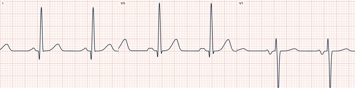
An inherited thickening of the heart muscle, usually the septum. The ECG often shows high voltage, deep narrow Q waves in the lateral and inferior leads and left atrial enlargement.
What the heart is doing
The thickened septum generates large septal forces, writing narrow, deep Q waves in leads that face away from it. The extra muscle raises QRS voltage, and the stiff ventricle enlarges the left atrium.
Why it looks this way
This is hypertrophic cardiomyopathy because high QRS voltage comes with deep, narrow Q waves in the lateral and inferior leads and a left atrial P wave.
Why it matters
A leading cause of sudden cardiac death in the young and in athletes. Septal Q waves in a young person with exertional symptoms need an echocardiogram. Outflow obstruction worsens with dehydration and vasodilators.
What to look for
- Deep, narrow “dagger” Q waves in lateral or inferior leads in a young person.
- High voltage with a broad, notched P in II or a deep negative P in V1.
- Ask about exertional syncope, chest pain and sudden death in the family.
Frequently asked questions
What are the ECG criteria for hypertrophic cardiomyopathy (septal)?
Voltage: often meets LVH criteria. Q waves: deep and narrow (< 40 ms) in lateral or inferior leads. ST and T: often lateral ST depression or T-wave inversion. P waves: left atrial enlargement. ECG: abnormal in most affected patients.
How is hypertrophic cardiomyopathy (septal) identified on an ECG?
This is hypertrophic cardiomyopathy because high QRS voltage comes with deep, narrow Q waves in the lateral and inferior leads and a left atrial P wave.
What can hypertrophic cardiomyopathy (septal) be mistaken for?
Prior inferior infarction: broader Q waves (≥ 40 ms) in one coronary territory. Left ventricular hypertrophy: high voltage without septal Q waves. Apical hypertrophic cardiomyopathy: giant T inversion in V3–V5.
What causes hypertrophic cardiomyopathy (septal)?
Common causes: sarcomere protein gene variants (autosomal dominant); family history of HCM or sudden death; and storage and infiltrative diseases that mimic it.