
Chamber enlargement
Apical hypertrophic cardiomyopathy
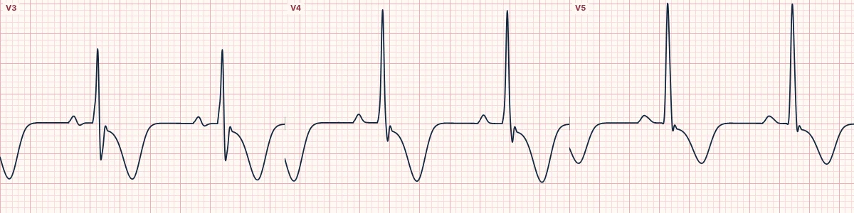
A form of hypertrophic cardiomyopathy in which the thickening is concentrated at the tip of the left ventricle, producing giant T-wave inversions in the chest leads.
What the heart is doing
Thick apical muscle generates large forces toward the mid-precordial leads and repolarizes in reverse order, so the T-wave vector points away from the apex and inverts deeply.
Why it looks this way
This is apical hypertrophic cardiomyopathy because high precordial voltage is combined with giant symmetric T-wave inversions in the mid-chest leads.
Why it matters
Generally milder than other forms of hypertrophic cardiomyopathy but can cause atrial fibrillation, apical aneurysm and ventricular arrhythmias. It imitates ischemia: confirm with imaging.
What to look for
- Very tall R waves in V4–V5 sitting above very deep, symmetric T inversions.
- The pattern is stable from one ECG to the next, unlike evolving ischemia.
- No Q waves and no loss of R waves.
Frequently asked questions
What are the ECG criteria for apical hypertrophic cardiomyopathy?
T waves: deep, symmetric inversion (classically ≥ 10 mm) in V3–V5. QRS: high voltage in the precordial leads. ST segment: mild depression in the same leads. Leads: mid-precordial, often lateral and inferior too.
How is apical hypertrophic cardiomyopathy identified on an ECG?
This is apical hypertrophic cardiomyopathy because high precordial voltage is combined with giant symmetric T-wave inversions in the mid-chest leads.
What can apical hypertrophic cardiomyopathy be mistaken for?
Wellens pattern type B: normal voltage, recent chest pain, changes evolve. Raised intracranial pressure: broad T inversions with a very long QT. LVH with strain: asymmetric, shallower T inversion in lateral leads.
What causes apical hypertrophic cardiomyopathy?
Common causes: inherited sarcomere gene variants; family history of cardiomyopathy; more prevalent in East Asian populations; and often found incidentally in adults.