
Chamber enlargement
Left atrial enlargement
An enlarged left atrium takes longer to depolarize, so the P wave becomes wide and notched in lead II, with a deep negative tail in V1.
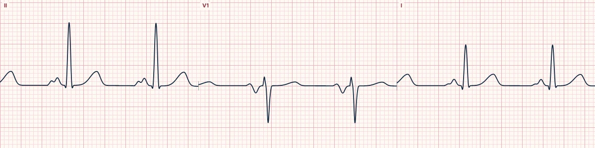
What the heart is doing
The right atrium depolarizes first, the left atrium second. A larger left atrium prolongs and enlarges the second component, which points leftward and backward, away from V1.
Why it looks this way
This is left atrial enlargement because the P wave in II is at least 120 ms wide and notched, with a deep terminal negative deflection in V1.
Why it matters
Suggests raised left-sided filling pressures and predicts atrial fibrillation. It is a clue to underlying valve or hypertensive disease and is not treated directly.
What to look for
- In II, the P wave is three small boxes wide or more, with two humps.
- In V1, the second half of the P wave dips at least one small box deep and wide.
- The QRS and PR can be entirely normal.
Frequently asked questions
What are the ECG criteria for left atrial enlargement?
P wave in II: ≥ 120 ms wide, notched ('M' shaped). Notch: peaks ≥ 40 ms apart. P wave in V1: terminal negative part ≥ 1 mm deep and ≥ 40 ms wide. P amplitude: normal (≤ 2.5 mm).
How is left atrial enlargement identified on an ECG?
This is left atrial enlargement because the P wave in II is at least 120 ms wide and notched, with a deep terminal negative deflection in V1.
What can left atrial enlargement be mistaken for?
Interatrial conduction delay: the same wide P wave with a normal-sized atrium. Ectopic atrial rhythm: abnormal P axis, not just a wide P wave. Right atrial enlargement: tall, narrow, peaked P waves.
What causes left atrial enlargement?
Common causes: mitral stenosis or regurgitation; hypertension and left ventricular hypertrophy; aortic stenosis; heart failure; and hypertrophic cardiomyopathy.