
The waves
The U wave
A small wave after the T
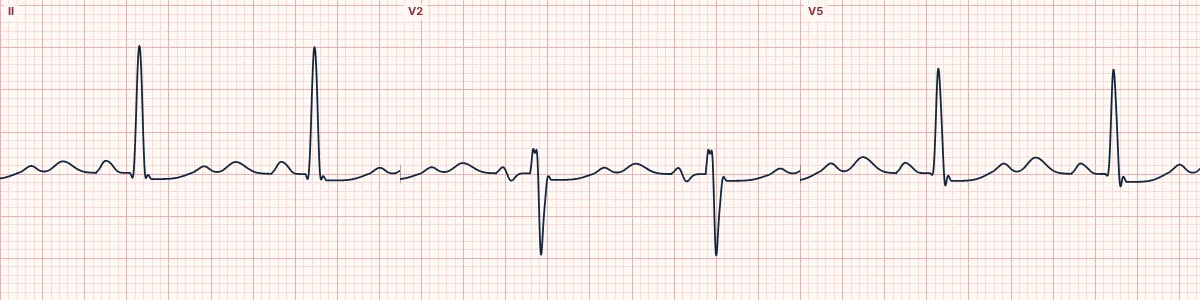
A small positive wave after the T wave, best seen in V2 and V3 at slow rates. Prominent U waves are a classic sign of low potassium.
What it is
Its origin is still debated: delayed repolarization of the Purkinje fibres or mid-myocardial cells, or afterpotentials from the mechanical stretch of the ventricles. It normally points the same way as the T wave and is about a tenth of its height.
When it grows
Low potassium, bradycardia and some drugs make the U wave larger. In hypokalemia the T wave flattens and the U wave grows until they merge, making the QT look long when it is really a QU interval.
Inverted U waves
A negative U wave in the chest leads is uncommon and points toward ischemia, hypertension or valve disease.
How to read it
- Look just after the T wave in V2 and V3.
- Do not include the U wave in the QT measurement.
Pitfalls
- A P wave of the next beat can look like a U wave at fast rates.
Frequently asked questions
The U wave: what is the key idea?
A small positive wave after the T wave, best seen in V2 and V3 at slow rates. Prominent U waves are a classic sign of low potassium.
The U wave: what are the key numbers?
Best seen: V2–V3; Normal size: < 1–1.5 mm, about 10% of T; Prominent: Hypokalemia, bradycardia, drugs.
The U wave: how do you apply it to an ECG?
Look just after the T wave in V2 and V3. Do not include the U wave in the QT measurement.
The U wave: what are the common pitfalls?
A P wave of the next beat can look like a U wave at fast rates.