
Heart muscle and systemic disease
Draft — pending owner review
COPD and emphysema
A heart seen through air
Hyperinflated lungs push the heart down and insulate it. The ECG shows low voltage, a rightward axis, tall P waves in the inferior leads and poor R-wave progression, with right heart strain in advanced disease.
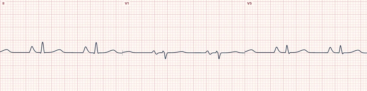
Hyperinflation
Air between the heart and the electrodes lowers voltage, especially in the left chest leads. The flattened diaphragm lets the heart hang vertically and rotate clockwise, giving a rightward axis, a late transition and sometimes S waves in I, II and III.
Right heart features
Pulmonary hypertension adds tall P waves (over 2.5 mm in II), a dominant R or rSR′ in V1 and right ventricular strain.
Rhythms
Multifocal atrial tachycardia and atrial fibrillation are common in exacerbations, often driven by low oxygen and bronchodilator drugs.
About the demonstration
The tracing combines low voltage, right atrial enlargement and a vertical axis.
How to read it
- Check limb-lead voltage and the axis.
- Look at P-wave height in II.
- Read the rhythm carefully for MAT.
Pitfalls
- Poor R-wave progression may be misread as an old anterior infarct.
- Electrodes placed low over hyperinflated lungs change the pattern.
Frequently asked questions
COPD and emphysema: what is the key idea?
Hyperinflated lungs push the heart down and insulate it. The ECG shows low voltage, a rightward axis, tall P waves in the inferior leads and poor R-wave progression, with right heart strain in advanced disease.
COPD and emphysema: what are the key numbers?
Voltage: Low, especially V5–V6; Axis: Rightward, often vertical; P waves: Tall in II, III and aVF; Rhythms: Multifocal atrial tachycardia, AF.
COPD and emphysema: how do you apply it to an ECG?
Check limb-lead voltage and the axis. Look at P-wave height in II. Read the rhythm carefully for MAT.
COPD and emphysema: what are the common pitfalls?
Poor R-wave progression may be misread as an old anterior infarct. Electrodes placed low over hyperinflated lungs change the pattern.