
Waves, voltage and chambers
Draft — pending owner review
Epsilon wave
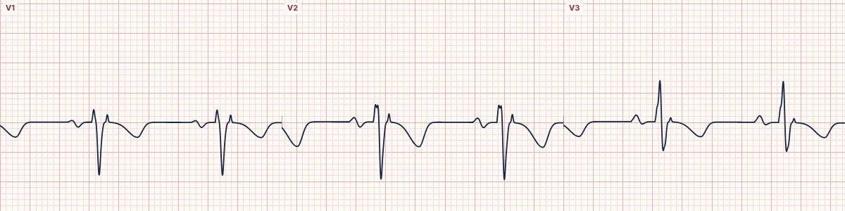
A late flicker after the QRS in V1–V3
A small, low-amplitude wiggle between the end of the QRS and the start of the T wave in V1–V3. It marks late activation of the right ventricle and is a diagnostic criterion for arrhythmogenic right ventricular cardiomyopathy.
What it looks like
A low, sharp notch or a few small waves just after the QRS, best seen in V1 and V2. It is easy to miss at standard settings; higher gain, faster paper speed or bipolar leads placed along the sternum (Fontaine leads) make it clearer.
Why it happens
In ARVC, fibrofatty tissue replaces right ventricular muscle and leaves islands of surviving cells that activate late, after the rest of the ventricles. Their small, delayed signal appears after the QRS in the leads over the right ventricle.
Weight as a criterion
It was a major criterion in the 2010 Task Force criteria; newer criteria give it less weight because observers often disagree about whether it is there. Rarely it appears after right ventricular infarction, in sarcoidosis or after repaired tetralogy of Fallot.
Companions
T-wave inversion in V1–V3 in adults without RBBB, a terminal activation duration of 55 ms or more in V1–V3, and PVCs or VT with an LBBB shape.
How to read it
- Pause, magnify V1–V3 and inspect the very start of the ST segment.
- Increase the gain or paper speed if your machine allows.
- Look for T inversion in V1–V3 and LBBB-shaped ectopy.
Pitfalls
- Noise and a small r′ are often over-read as epsilon waves.
- A normal ECG does not exclude ARVC, especially early on.
Frequently asked questions
Epsilon wave: what is the key idea?
A small, low-amplitude wiggle between the end of the QRS and the start of the T wave in V1–V3. It marks late activation of the right ventricle and is a diagnostic criterion for arrhythmogenic right ventricular cardiomyopathy.
Epsilon wave: what are the key numbers?
Leads: V1–V3; Timing: After the QRS, before the T wave; Terminal activation duration: ≥ 55 ms in V1–V3 is a related criterion; Disease: Arrhythmogenic right ventricular cardiomyopathy.
Epsilon wave: how do you apply it to an ECG?
Pause, magnify V1–V3 and inspect the very start of the ST segment. Increase the gain or paper speed if your machine allows. Look for T inversion in V1–V3 and LBBB-shaped ectopy.
Epsilon wave: what are the common pitfalls?
Noise and a small r′ are often over-read as epsilon waves. A normal ECG does not exclude ARVC, especially early on.