
Waves, voltage and chambers
Draft — pending owner review
R-wave peak time (intrinsicoid deflection)
How long activation takes to reach the surface
The time from the start of the QRS to the peak of the R wave. Long in the left lateral leads it supports LVH, LBBB or LAFB; in lead II during a wide tachycardia, 50 ms or more favors VT.
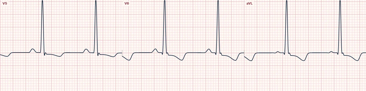
Measuring it
Mark the start of the QRS and the top of the R wave (the last R wave if there is an R′) in the same lead. Normal values are up to about 35 ms in V1–V2 and about 45 ms in V5–V6 and aVL.
What prolongs it
Anything that slows activation of the wall under the electrode: a thick left ventricle (LVH), loss of the left bundle (LBBB) or of the anterior fascicle (LAFB, best seen in aVL). In V1–V2 it lengthens with RBBB and RVH.
In wide-complex tachycardia
Measured in lead II from the start of the QRS to its first change in direction, whether the QRS is positive or negative, a value of 50 ms or more favors VT, because activation that starts in muscle is slow from the very beginning.
How to read it
- Use a lead with a sharp QRS onset.
- Measure with the calipers on the paused tracing.
- Judge the value against the lead you measured in.
Pitfalls
- The QRS onset is hard to see in noisy tracings.
- In LBBB and fascicular block it is long by definition and adds little.
Frequently asked questions
R-wave peak time (intrinsicoid deflection): what is the key idea?
The time from the start of the QRS to the peak of the R wave. Long in the left lateral leads it supports LVH, LBBB or LAFB; in lead II during a wide tachycardia, 50 ms or more favors VT.
R-wave peak time (intrinsicoid deflection): what are the key numbers?
V1–V2: Normal up to about 35 ms; V5–V6 and aVL: Normal up to about 45 ms; Lead II in a wide tachycardia: ≥ 50 ms favors VT.
R-wave peak time (intrinsicoid deflection): how do you apply it to an ECG?
Use a lead with a sharp QRS onset. Measure with the calipers on the paused tracing. Judge the value against the lead you measured in.
R-wave peak time (intrinsicoid deflection): what are the common pitfalls?
The QRS onset is hard to see in noisy tracings. In LBBB and fascicular block it is long by definition and adds little.