
Approach to tachycardia
Wide-complex tachycardia
VT until proven otherwise
A wide QRS at a fast rate is ventricular tachycardia in most adults, especially those with structural heart disease. A few features make VT almost certain; none makes it safe to assume the opposite.
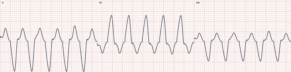
Features that prove VT
AV dissociation: P waves marching through at their own slower rate. Capture beats: a normal narrow beat when a sinus impulse gets through. Fusion beats: a hybrid of the two. Any of these confirms a ventricular origin.
Features that favour VT
QRS wider than 160 ms. Extreme (northwest) axis. Concordance: all chest leads pointing the same way. No RS complex in any chest lead, or an RS interval longer than 100 ms. History of infarction, cardiomyopathy or heart failure.
The alternatives
Supraventricular tachycardia with bundle branch block (pre-existing or rate-related), antidromic AV re-entry, pre-excited atrial fibrillation (irregular, very fast and varying in width), hyperkalemia and sodium-channel blocker toxicity, and paced rhythms.
Practical rule
When uncertain, treat as ventricular tachycardia. Drugs that block the AV node can cause collapse in VT and in pre-excited atrial fibrillation.
How to read it
- Check the patient first: pulse, blood pressure, perfusion.
- Search every lead for P waves independent of the QRS.
- Look for capture and fusion beats, axis and concordance.
Pitfalls
- Good blood pressure does not make the rhythm supraventricular.
- Irregular, bizarre and very fast wide complexes may be pre-excited atrial fibrillation: avoid AV-nodal blockers.
Frequently asked questions
Wide-complex tachycardia: what is the key idea?
A wide QRS at a fast rate is ventricular tachycardia in most adults, especially those with structural heart disease. A few features make VT almost certain; none makes it safe to assume the opposite.
Wide-complex tachycardia: what are the key numbers?
Proves VT: AV dissociation, capture, fusion beats; Favours VT: QRS > 160 ms, extreme axis, concordance; Irregular and wide: Consider pre-excited AF or polymorphic VT.
Wide-complex tachycardia: how do you apply it to an ECG?
Check the patient first: pulse, blood pressure, perfusion. Search every lead for P waves independent of the QRS. Look for capture and fusion beats, axis and concordance.
Wide-complex tachycardia: what are the common pitfalls?
Good blood pressure does not make the rhythm supraventricular. Irregular, bizarre and very fast wide complexes may be pre-excited atrial fibrillation: avoid AV-nodal blockers.