
Wide-complex tachycardia criteria
Draft — pending owner review
Josephson sign
A notch near the bottom of the S wave in VT
Notching or slurring near the nadir of the S wave, usually in V1–V2, during a wide-complex tachycardia. It reflects slow, uneven muscle-to-muscle conduction and favors VT over SVT with aberrancy.
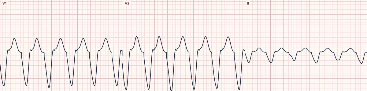
The sign
In a wide-complex tachycardia with an LBBB-like shape, look at the S wave in V1 and V2. A notch or slur on the way down to the deepest point of the S wave, or close to it, is Josephson's sign.
Why it happens
Aberrant conduction still uses the Purkinje network on the unblocked side, so the first part of the QRS is fast and smooth. A ventricular focus spreads through muscle, often unevenly around scar, and the wavefront's hesitations show up as notches.
Using it
It is one of several morphology clues and none is decisive alone. In V1–V2 it sits alongside a broad initial R wave (over 30 ms) and a long interval to the S nadir (over 60 ms) as features of VT with an LBBB shape.
About the demonstration
The tracing is the lab's LBBB-morphology VT, drawn from a smooth model of activation, so its S waves are not notched. Use it to find where the sign would appear; in real VT, check every chest lead.
How to read it
- Pause the tracing and magnify V1 and V2.
- Follow the downstroke of the S wave to its lowest point.
- Combine with AV dissociation and the algorithms before deciding.
Pitfalls
- Baseline noise can mimic a notch; check several beats.
- Hyperkalemia and drug toxicity also produce slurred, wide complexes.
Frequently asked questions
Josephson sign: what is the key idea?
Notching or slurring near the nadir of the S wave, usually in V1–V2, during a wide-complex tachycardia. It reflects slow, uneven muscle-to-muscle conduction and favors VT over SVT with aberrancy.
Josephson sign: what are the key numbers?
Companion clues in V1–V2: Initial R > 30 ms; onset to S nadir > 60 ms; Absence: Proves nothing.
Josephson sign: how do you apply it to an ECG?
Pause the tracing and magnify V1 and V2. Follow the downstroke of the S wave to its lowest point. Combine with AV dissociation and the algorithms before deciding.
Josephson sign: what are the common pitfalls?
Baseline noise can mimic a notch; check several beats. Hyperkalemia and drug toxicity also produce slurred, wide complexes.