
Rhythm clues and devices
Draft — pending owner review
Sinus node re-entrant tachycardia
Sinus-shaped P waves that start and stop suddenly
A re-entrant tachycardia within or around the sinus node. The P waves look exactly like sinus P waves, but the rhythm starts and stops abruptly, often after a premature atrial beat, usually at 100–150/min.
How it differs from sinus tachycardia
Sinus tachycardia speeds up and slows down gradually with need. Sinus node re-entry switches on and off suddenly, often triggered by a premature atrial complex, and can be stopped by vagal maneuvers or adenosine.
ECG
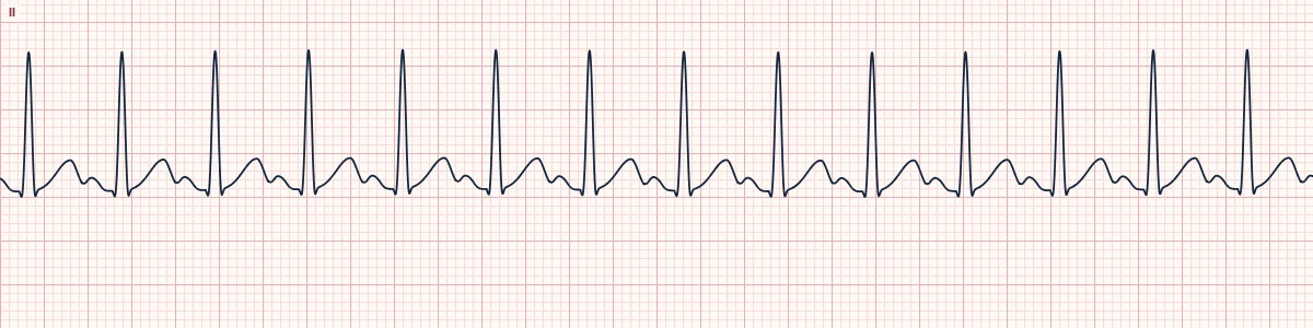
Sinus-shaped P waves (upright in II, inverted in aVR), a long RP interval, a rate usually of 100–150/min, and a PR that may lengthen a little at the faster rate.
Why it matters
It is uncommon and easily dismissed as anxiety-driven sinus tachycardia. Catching the onset or end on a monitor makes the diagnosis.
About the demonstration
The tracing shows sinus-shaped P waves at 140/min, as during an episode; a short strip cannot show the sudden start and stop.
How to read it
- Find the onset or end on a monitor strip.
- Compare the P waves with the patient's sinus P waves.
- Look for a triggering premature atrial beat.
Pitfalls
- Without the onset or end, it cannot be told from sinus tachycardia.
Frequently asked questions
Sinus node re-entrant tachycardia: what is the key idea?
A re-entrant tachycardia within or around the sinus node. The P waves look exactly like sinus P waves, but the rhythm starts and stops abruptly, often after a premature atrial beat, usually at 100–150/min.
Sinus node re-entrant tachycardia: what are the key numbers?
Rate: Usually 100–150/min; P waves: Identical to sinus P waves; Onset: Abrupt, often after a PAC; Response: Often stops with vagal maneuvers or adenosine.
Sinus node re-entrant tachycardia: how do you apply it to an ECG?
Find the onset or end on a monitor strip. Compare the P waves with the patient's sinus P waves. Look for a triggering premature atrial beat.
Sinus node re-entrant tachycardia: what are the common pitfalls?
Without the onset or end, it cannot be told from sinus tachycardia.