
Recording problems
Draft — pending owner review
V1 and V2 placed too high
Electrodes in the second or third rib space
Placing V1 and V2 one or two rib spaces too high produces an rSr′ pattern, negative or biphasic P waves and T inversion in those leads. It mimics incomplete RBBB, a Brugada type 2 pattern or an old anterior infarct.
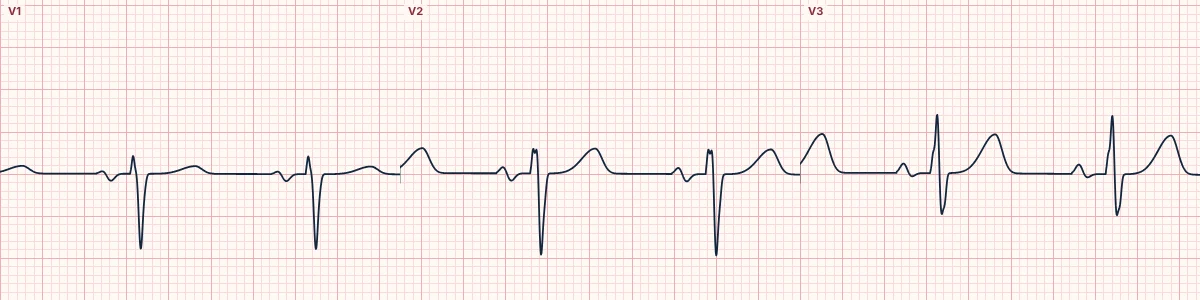
What changes
Higher electrodes look down on the heart from above: the P wave in V1–V2 becomes negative or biphasic (like aVR), an r′ appears, R waves shrink and T waves may invert.
How to spot it
A negative or deeply biphasic P wave in V2 suggests high placement, because with correct placement the P in V2 is usually upright. Comparing with V3 and with an old ECG helps.
Why it matters
False RBBB, Brugada or infarct diagnoses lead to needless tests. Conversely, recording V1–V2 deliberately one and two spaces higher is used to unmask a type 1 Brugada pattern.
About the demonstration
The lab always places V1 and V2 correctly, so the tracing shows the normal appearance to compare against: a small r wave, a deep S wave and an upright or small biphasic P wave.
How to read it
- Count down from the angle of Louis (second rib) to find the fourth space.
- Check the P wave in V1 and V2.
- Repeat the ECG with correct placement if in doubt.
Pitfalls
- Electrodes are often placed too high on women and on large chests.
Frequently asked questions
V1 and V2 placed too high: what is the key idea?
Placing V1 and V2 one or two rib spaces too high produces an rSr′ pattern, negative or biphasic P waves and T inversion in those leads. It mimics incomplete RBBB, a Brugada type 2 pattern or an old anterior infarct.
V1 and V2 placed too high: what are the key numbers?
Correct V1: 4th intercostal space, right sternal edge; Correct V2: 4th intercostal space, left sternal edge; Clue to high placement: Negative P wave in V2; Mimics: Incomplete RBBB, Brugada type 2, old anterior MI.
V1 and V2 placed too high: how do you apply it to an ECG?
Count down from the angle of Louis (second rib) to find the fourth space. Check the P wave in V1 and V2. Repeat the ECG with correct placement if in doubt.
V1 and V2 placed too high: what are the common pitfalls?
Electrodes are often placed too high on women and on large chests.