
Waves, voltage and chambers
Draft — pending owner review
Biatrial enlargement
Both atria at once
Signs of right and left atrial enlargement on the same ECG: a P wave in II that is both tall and broad, or a biphasic P in V1 with a tall first part and a deep, wide second part.
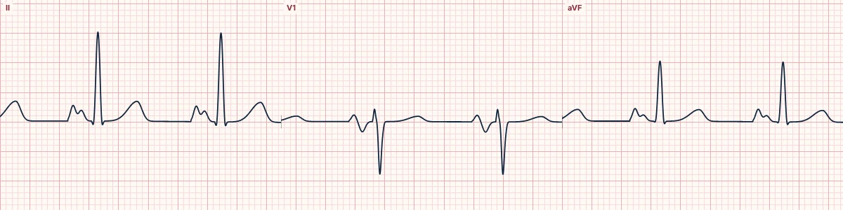
Putting the two signs together
Right atrial enlargement makes the first part of the P wave taller; left atrial enlargement makes the second part later, broader and, in V1, deeply negative. When both are enlarged, the P wave is tall and wide in II and both halves of the P in V1 are exaggerated.
Causes
Mitral valve disease with pulmonary hypertension, congenital heart disease, dilated or restrictive cardiomyopathy, and long-standing lung disease with left heart disease.
Limits
P-wave criteria are insensitive and depend on rhythm and lead placement. The echocardiogram, not the ECG, sizes the atria.
How to read it
- Measure P height and width in II.
- Look at the two halves of the P wave in V1.
- Make sure the rhythm is sinus before reading P size.
Pitfalls
- V1 placed too high distorts the P wave.
- A notched P in II is not enough without the width.
Frequently asked questions
Biatrial enlargement: what is the key idea?
Signs of right and left atrial enlargement on the same ECG: a P wave in II that is both tall and broad, or a biphasic P in V1 with a tall first part and a deep, wide second part.
Biatrial enlargement: what are the key numbers?
Right atrial: P > 2.5 mm in II, or first part of P > 1.5 mm in V1; Left atrial: P ≥ 120 ms in II; terminal P in V1 ≥ 1 mm and ≥ 40 ms; Biatrial: Features of both.
Biatrial enlargement: how do you apply it to an ECG?
Measure P height and width in II. Look at the two halves of the P wave in V1. Make sure the rhythm is sinus before reading P size.
Biatrial enlargement: what are the common pitfalls?
V1 placed too high distorts the P wave. A notched P in II is not enough without the width.