
Ventricular
Draft — pending owner review
Fascicular ventricular tachycardia
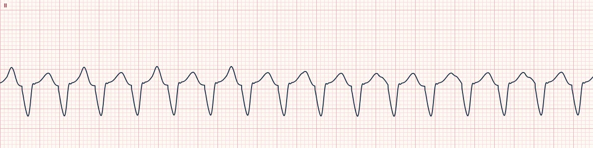
An idiopathic VT from the left posterior fascicle, usually in young adults with normal hearts. The QRS is only moderately wide, with an RBBB shape and left axis deviation.
What the heart is doing
A re-entrant circuit runs through Purkinje tissue around the left posterior fascicle. It exits into the conduction system near the inferior septum, so activation spreads quickly and the QRS stays relatively narrow.
Why it looks this way
This is fascicular VT because a regular tachycardia with a QRS of about 130 ms has an RBBB shape with left axis deviation, dissociated from the P waves.
Why it matters
Easily mistaken for SVT with aberrancy because the QRS is only modestly wide and the patient is young and well. Ablation is usually curative. Any regular wide tachycardia is still treated as VT until proven otherwise.
What to look for
- A regular, modestly wide tachycardia shaped like RBBB plus left anterior fascicular block.
- AV dissociation or fusion beats, which prove VT.
- A young, well patient with no known heart disease.
Frequently asked questions
What are the ECG criteria for fascicular ventricular tachycardia?
QRS: about 120–140 ms, RBBB-like in V1. Axis: left axis deviation (posterior fascicle origin). RS interval: relatively short, often under 80 ms. AV relationship: dissociation or fusion beats when looked for. Response: often stops with verapamil; adenosine usually does not.
How is fascicular ventricular tachycardia identified on an ECG?
This is fascicular VT because a regular tachycardia with a QRS of about 130 ms has an RBBB shape with left axis deviation, dissociated from the P waves.
What can fascicular ventricular tachycardia be mistaken for?
Bifascicular block: the same shape in sinus rhythm, with a P wave before every QRS. SVT with RBBB aberrancy: P waves linked to every QRS. Monomorphic VT: usually wider, in a heart with scar or cardiomyopathy.
What causes fascicular ventricular tachycardia?
Common causes: re-entry within the left posterior fascicle (most common); left anterior fascicle origin (rarer, right axis); exercise or emotional stress; and usually no structural heart disease.