
Ischemia & prior infarction
Draft — pending owner review
Left ventricular aneurysm (persistent ST elevation)
ST elevation that persists weeks after an anterior infarction, sitting over Q or QS waves, caused by a scarred, bulging segment of ventricle. It is easily mistaken for an acute STEMI.
What the heart is doing
The thin, akinetic scar no longer depolarizes, leaving Q waves. Its abnormal wall tension and motion keep an injury-like current flowing, so the ST segment stays elevated long after the infarct.
Why it looks this way
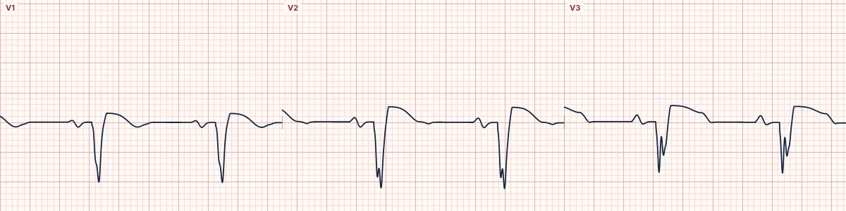
This is a left ventricular aneurysm pattern because convex ST elevation sits over QS waves in V1–V3 while the T waves stay small relative to the QRS.
Why it matters
The danger is confusing it with a new STEMI, or the reverse; old ECGs and the T/QRS ratio help, and a new occlusion can still occur. Aneurysms predispose to heart failure, mural thrombus and ventricular tachycardia.
What to look for
- Find the Q or QS waves underneath the ST elevation.
- Divide T-wave height by QRS amplitude in V1–V4: small T waves point to an old aneurysm.
- Compare with old ECGs: unchanged ST elevation favors aneurysm.
Frequently asked questions
What are the ECG criteria for left ventricular aneurysm (persistent ST elevation)?
QRS: Q or QS waves in V1–V4. ST segment: persistent elevation, usually convex, two weeks or more after MI. T waves: small relative to the QRS. T/QRS ratio: below 0.36 in every lead V1–V4 favors aneurysm; above it in any lead suggests acute MI. Comparison: unchanged from earlier ECGs.
How is left ventricular aneurysm (persistent ST elevation) identified on an ECG?
This is a left ventricular aneurysm pattern because convex ST elevation sits over QS waves in V1–V3 while the T waves stay small relative to the QRS.
What can left ventricular aneurysm (persistent ST elevation) be mistaken for?
Mid LAD occlusion: acute, with T waves large relative to the QRS. Prior anterior infarction: Q waves without persistent ST elevation. Benign early repolarization: no Q waves, and tall T waves.
What causes left ventricular aneurysm (persistent ST elevation)?
Common causes: prior large anterior infarction; late or no reperfusion of the infarct artery; and prior inferior infarction (inferior aneurysm, less common).